

The dedicated Pawar Multispeciality Hospital is one of the best hospitals in Pune for Echo Cardiographs. The hospital conducts various Echo Cardiographs.
The heart is a two-stage electrical pump that circulates blood throughout the body. The anatomy includes four chambers and four valves. For the heart to function normally these structures need to be intact and the heart muscle needs to beat in a coordinated fashion, so that blood flows in and out of each chamber in the proper direction.
An echocardiogram (echo=sound + card=heart + gram=drawing) is an ultrasound test that can evaluate the structures of the heart, as well as the direction of blood flow within it. Technicians specially trained in echocardiography produce the images and videos, often using a special probe or transducer that is placed in various places on the chest wall, to view the heart from different directions. Cardiologists, or heart specialists, are trained to evaluate these images to assess heart function and provide a report of the results.The echocardiogram is just one of the many tests that can be done to evaluate heart anatomy and function.
An electrocardiogram (EKG, ECG) is the most common heart tracing done. Electrodes are placed on the chest wall and collect information about the electrical activity of the heart. Aside from the rate and rhythm of the heartbeat, the EKG can provide indirect evidence of blood flow within arteries to heart muscle and the thickness of heart muscle.

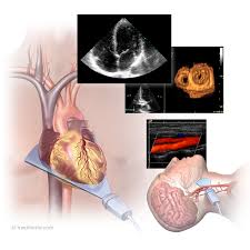
Transthoracic echocardiogram: In the transthoracic echocardiogram procedure, the echocardiographer places the transducer, or probe, on the chest wall and bounces sound waves off the structures of the heart. The return signals are received by the same transducer and converted by a computer into the images seen on the screen.
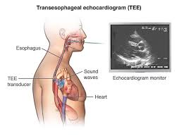
Transesophageal echocardiogram: In some situations, a clearer view of the heart is required and instead of placing the transducer on the chest wall, a cardiologist will direct the probe through the mouth into the esophagus. The esophagus is located right next to the heart in the middle of the chest and the sound waves can travel to the heart without the interference of the ribs and muscles of the chest wall.
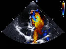
Doppler echocardiogram: In addition to sound waves bouncing off the solid structures of the heart, they also bounce off the red blood cells as they circulate through the heart chambers. Using Doppler technology, the echocardiogram can assess the speed and direction of blood flow, helping increase the amount and quality of information available from the test. The computer can add color to help the doctor appreciate that information. Color flow Doppler is routinely added to all echocardiogram studies and is the same technology used in weather reports.
Stress echocardiogram: A stress echocardiogram helps uncover abnormalities in the function of the heart wall muscle. The patient may be asked to exercise by either walking on a treadmill or riding an exercise bicycle. The echocardiogram is performed before exercise as a baseline, and then immediately after the test.
When coronary arteries narrow due to atherosclerotic heart disease, the heart muscle may not receive enough blood supply during exercise. During a stress echocardiogram, the areas of heart muscle not receiving enough blood flow may not squeeze as well as other parts of the heart, and will appear to have motion abnormality. This can indirectly indicate narrowing, or stenosis, of the coronary arteries. This can cause chest pain (angina), shortness of breath, or you may have no symptoms.
For a stress echocardiogram to be effectively interpreted, the exercise done needs to achieve certain minimum intensity. If the patient is unable to exercise adequately, medications can be injected intravenously to chemically make the heart respond as if exercise is occurring.
Contrast may be injected into the patient's vein to help enhance the images and increase the information that is obtained. The contrast material (Optison, Density) are microscopic protein shells filled with gas bubbles. The decision to use contrast depends upon the patient's specific situation.
The purpose of an echocardiogram is to assess the structure and function of the heart. It is recommended as a non-invasive procedure as part of assessing potential and established heart problems.
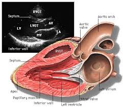
Regarding structure, the test can assess the general size of the heart, the size of the four heart chambers, and the appearance and function of the four heart valves. It can look at the two septa of the heart; the atrial septum separates the right and left atrium and the ventricular septum separates the right and left ventricles. It can also assess the pericardium (the sac that lines the heart) and the aorta.
Regarding function, the echocardiogram can determine how the heart valves open and close. It can evaluate whether the heart muscle squeezes appropriately and how efficiently. Cardiac output measures how much blood the heart pumps. Ejection fraction measures what percent of blood within the heart is pumped out to the body with each heartbeat. It can also measure how well the heart relaxes in between beats, when the heart fills for the next pump.
Some heart issues that the echocardiogram can help evaluate include the following:
Heart valve disorders: Stenosis (narrowed), insufficiency or regurgitation (leaking), and endocarditis (infection of the valves)
Abnormalities of the septum: Atrial septal defect, ventricular septal defect, and patent foramen ovale
Wall motion abnormalities: Cardiomyopathy, atherosclerotic heart disease (also known as coronary artery disease), and trauma
Diseases of the pericardium (the sac that lines the heart): This includes pericardial effusion (assessment of fluid in the pericardial sac)

There is no preparation for a transthoracic echocardiogram.
When a transesophageal echocardiogram is performed, the patient usually requires some sedation to tolerate the procedure. The stomach should be empty to prevent vomiting and aspiration into the lungs. For that reason, the patient should have nothing to eat or drink for eight hours before the procedure. Due to the sedation, the patient will need a family member or friend to escort the patient home.
For a stress echocardiogram, the patient may need to walk on a treadmill or ride a bicycle. Comfortable shoes are recommended.

An echocardiogram is an office or outpatient procedure. Electrodes are placed on the chest wall to monitor heart rate and rhythm. The lights in the room may be dimmed to help see the images on the computer monitor. If contrast is used, an intravenous line will need to be started.
In transthoracic echocardiogram, the patient's chest will need to be exposed. The technician will press the transducer or probe firmly on the chest wall to get the heart images. The patient may be asked to roll on their left side take deep breaths to help the probe better "see" the heart. The patient will be monitored because of the need for intravenous sedation. A heart monitor and oxygen monitor will be placed; supplemental oxygen is usually provided by prongs placed in the nose and an intravenous line will be started. Once sedated, the cardiologist will pass a tube, with the transducer on its tip, through the mouth and position it in the esophagus at a level near the heart. The patient may or may or remember the procedure because many of the sedative medicines have an amnestic effect; but once the patient is fully awake, they may be discharged home with an escort.